"The course provides basic introductory information to Sonographers who are new to Pelvic Sonography."
Course Participant
-
CPD points (SSR) – Pending approval
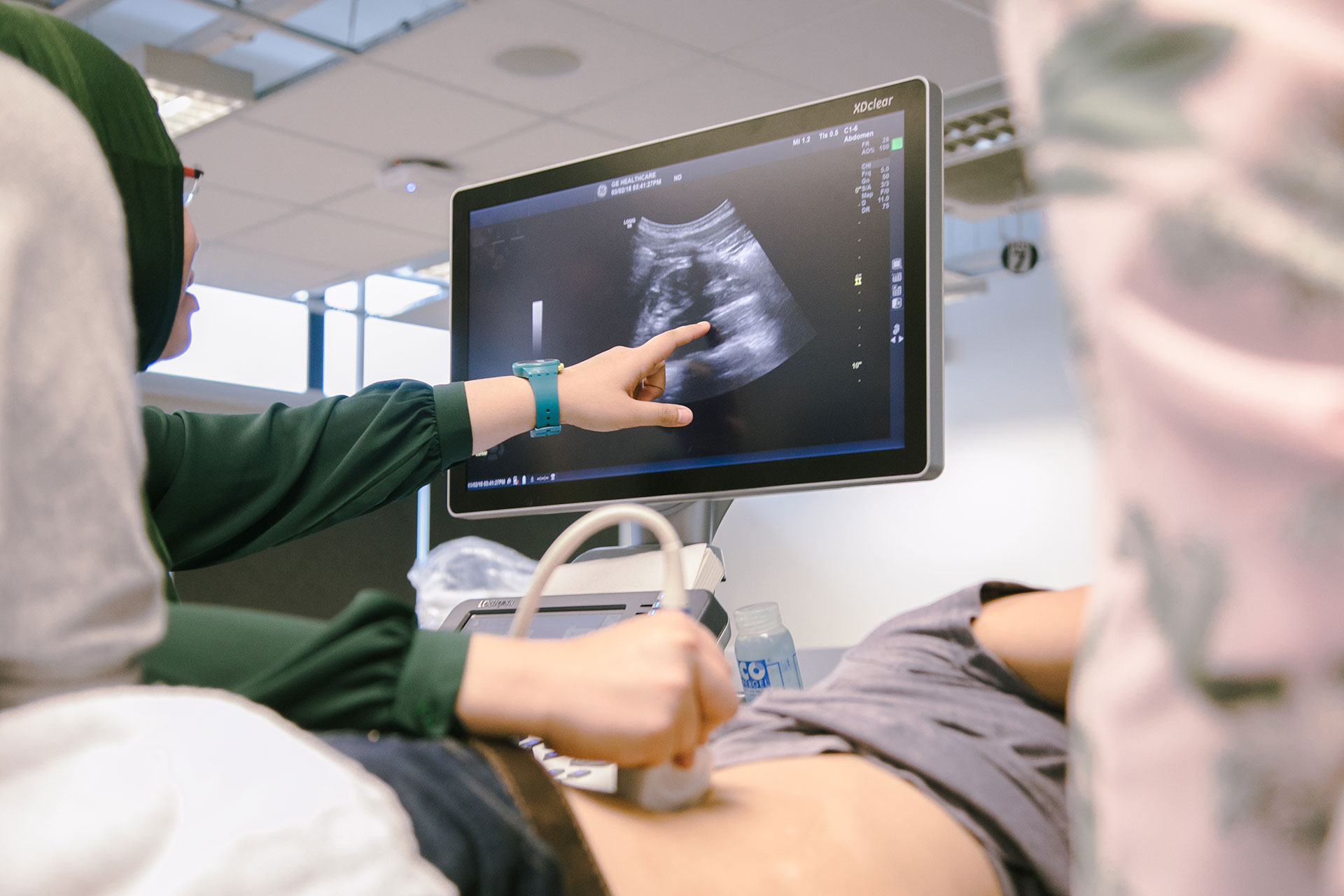
Ultrasound plays a crucial role in the evaluation and management of patients with pelvic diseases and injuries.
The workshop will provide participants with the underpinning academic knowledge and clinical skills needed to undertake a range of gynaecological ultrasound examinations.
It will focus on indications, anatomy, scanning techniques/protocol, and normal and abnormal findings involved in a pelvic ultrasound examination.
The topics for the male pelvis will include common indications for ultrasound, gross and sonographic anatomy of the prostate, sonographic anatomy, scanning techniques, normal and abnormal findings of the prostate and seminal vesicles, and technical pitfalls of prostate sonography.
The topics for the female pelvis include gross and sonographic anatomy of the female pelvis, menstrual phases and relevant imaging appearances, scanning protocols, techniques, and common pathologies as well as technical pitfalls associated with female pelvic sonography. Groin, abdominal wall, and bowel sonography will also be covered in the workshop.
Hands-on practical will be conducted in the afternoon sessions to supplement the didactic teaching. It is ideal for practitioners who wish to expand their scope of practice in pelvic sonography and can be taken as a single module for continuing professional development.

Senior Lecturer, Medical Ultrasound, University of The West of England, Bristol UK (UWE), Advanced Practitioner, Ultrasound Department, Royal United Hospital Foundation Trust (RUH) Bath UK
| Run | Dates | Delivery and Time |
|---|---|---|
| Sep 2025 run | 17 – 28 Sep 2025 | Asynchronous online |
| 29 – 30 Sep 2025 | In-person 8:30 am – 5:00 pm |
| Time | Topics |
|---|---|
| 8:30 am | Welcome and registration |
| 9:00 am | Introduction to pelvic sonography Instrumentation, system presets and technical requirements |
| 9:30 am | The female pelvis |
| 10:30 am | Break |
| 11:00 am | The female pelvis Assessment of uterine myometrium/vagina/cervix |
| 12:30 pm | Lunch |
| 1:30 pm | The female pelvis Endometrial assessment |
| 2:30 pm | Practical hands-on session |
| 5:00 pm | End of Day 1 |
| Time | Topics |
|---|---|
| 8:30 am | Recap of Day 1 |
| 9:00 am | The female pelvis Assessment of ovarian and adnexal pathology |
| 10:30 am | Break |
| 11:00 am | The female pelvis Assessment of post-menopausal women, pelvic non gynaecological pathology/ retro uterine pouch |
| 12:30 pm | Lunch |
| 1:30 pm | Practical hands-on session |
| 5:00 pm | End of day 2 |
A Certificate of Participation will be issued to participants who
The full fee for this course is S$4,054.80.
| Category | After SF Funding |
|---|---|
| Singapore Citizen (Below 40) | S$1,216.44 |
| Singapore Citizen (40 & Above) | S$472.44 |
| Singapore PR / LTVP+ Holder | S$1,216.44 |
| Non-Singapore Citizen | S$4,054.80 (No Funding) |
Note: All fees above include GST. GST applies to individuals and Singapore-registered companies.
New Engineering Micro-credentials Launching Soon!
Exciting news! We are introducing new micro-credentials in Electrical and Electronic Engineering & Infrastructure and Systems Engineering. Be among the first to know by registering your interest today! Register now →