Overview
Preventive healthcare and early intervention have been proven to vastly reduce the risk of disease, disabilities, and death.
With the national shift toward these approaches in Singapore, the demand for specialists in sonography has naturally risen.
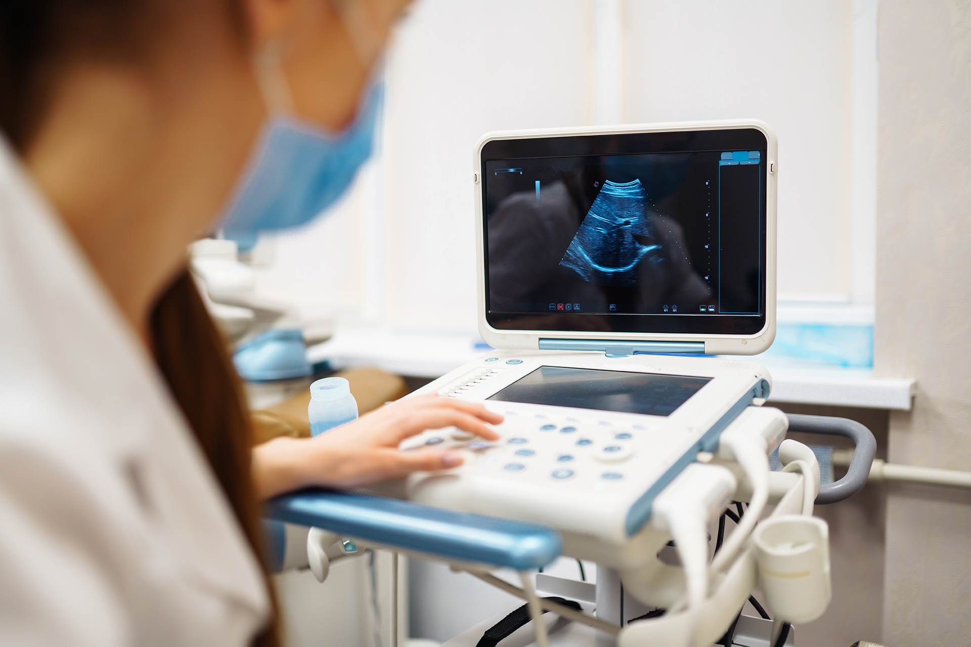
The SIT Graduate Certificate in Medical Sonography (GCert MS) is designed with a clinical focus to deepen and broaden the skills of allied health professionals, enabling them to apply scientific knowledge, specialist expertise and advanced practical skills to the clinical applications of ultrasound. It supports Singapore’s efforts to build a future-ready healthcare workforce, especially in the context of an ageing population facing chronic diseases that require accurate diagnosis.
Each module, worth 6 credits, can be taken individually or stack towards a Graduate Certificate in Medical Sonography.
Modules Available
Modules are stackable towards a GCert MS. They can also be taken individually.
Candidates will be awarded a GCert MS if they pass 3 modules totalling 18 credits (including 1 Core Module) within 2 years with a CGPA of ≥ 2.0., which can then be stacked to SIT's Master of Health Sciences.
Duration
Each module is around 12 – 14 weeks in duration. Candidates are expected to set aside time for self-directed learning for the entire duration of the module.
Prerequisites
- Relevant ultrasound-related experience with at least 1 year of working experience as an independent sonographer
- Have access to a medical ultrasound facility for clinical applications
- Submission of the following documents:
- Employment support letter (Includes applicant's area of work)*
- Recognised BSc degree in any of the health sciences registrable with the local regulatory bodies*
- Transcript of highest relevant qualification*
- Professional certification
- Answers to the following questions:
- Your reasons for pursuing the module (100 - 150 words)
- Why did you select SIT over other academic institutions? (100 - 150 words)
- What do you hope to achieve at the completion of the module? (100 - 150 words)
Note:
* Mandatory documents
Core Modules
You are required to complete either one of the Core Modules.
| Code | Module | Credits |
|---|---|---|
| HSC6002 | Evidence-based Practice and Ethics | 6 |
| HSC6003 | Health and Ageing | 6 |
Specialist Modules
You are required to complete the following two Specialist Modules.
| Code | Module | Credits |
|---|---|---|
| DRG6101 | Ultrasound Physics, Instrumentation & Quality Assurance | 6 |
| DRG6102 | Abdominal Ultrasound | 6 |
Note:
-
The above modules are subsidised by SkillsFuture Singapore.
-
Specialist Modules are known as Discipline Electives under SIT's Master of Health Sciences programme.
Certificate and Assessment
A Certificate of Attainment for each module will be issued to candidates who
- Attend at least 75% of the module; and
- Undertake and pass credit-bearing assessment during the module
Participants who meet the attendance requirement but do not pass the assessment will receive a Certificate of Participation.
Frequently Asked Questions
-
How does stacking to SIT's Master of Health Sciences work?
Our Graduate Certificate in Medical Sonography includes one core module and two specialist modules (also known as Discipline Electives). These modules are also components of the Master of Health Sciences programme. By completing the Graduate Certificate, you can transition into the Master of Health Sciences programme and apply these credits towards earning the MHSc degree.
-
Are the modules conducted full-time or part-time? Am I able to continue working if the module is full-time?
Both full-time and part-time modes are available.
For the part-time mode, each module will be delivered using blended learning pedagogy (hybrid of online learning and flexible workshop scheduling) for adult learners.
Candidates who are pursuing the full-time mode are required to have access to a medical ultrasound facility and clock in at least 3 days/week of clinical experience at the clinical site to fulfil the clinical requirement. This is important to develop the necessary technical, clinical, and diagnostic skills during the candidature.
Candidates undertaking the full-time mode can take more than one module (up to a maximum of 3 modules) in the same trimester. However, during the course of study, candidates who are pursuing the full-time mode are required to have adequate access to clinical work to fulfill the clinical development.
Candidates must complete logging the required number of clinical scans in the portfolio and develop competency in the respective clinical applications. They must be actively engaged in a supervised ultrasound clinical practice in the workplace.
At the end of the trimester, candidates must satisfy the clinical requirements (i.e., compulsory logging of the required number of ultrasound examinations in the clinical portfolio and passing the clinical skills assessment).
-
Is AHPC a compulsory requirement for Medical Sonography modules?
The AHPC requirement is preferred but not a compulsory entry criterion for the Graduate Certificate in Medical Sonography.
-
Am I allowed to take more modules concurrently to complete the course within a shorter time span?
It is possible to take all modules offered within the same trimester. The module timings are scheduled not to overlap.
Acceptance is granted at the sole discretion of SITLEARN.
-
How will the modules be conducted?
Classes are conducted in the day or in the evenings, after office hours. Additionally, candidates must allocate time for self-study, developing clinical portfolio, participating in online discussions, completing continual assessments, and preparing for a final exam.
-
How will the candidates be graded for the modules?
Each module may vary slightly, but all will include continual assessments followed by a final exam.
Explore More Courses
Designed for busy professionals and executives, our courses are designed for those with ambition to grow in their career. Discover new opportunities to explore and hone your craft with us.
New Engineering Micro-credentials Launching Soon!
Exciting news! We are introducing new micro-credentials in Electrical and Electronic Engineering & Infrastructure and Systems Engineering. Be among the first to know by registering your interest today! Register now →
